= (Scintillating Scotoma) =
Artist, Title, Date
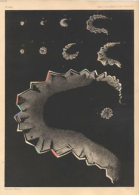
Hubert Airy, MD, Depiction of Scotomas in Transient Hemiopsia (commonly referred to as Scintillating Scotoma), 1870. Chromolithograph published in the Philosophical Transactions of the Royal Society of London.
Description of Disease & Etiology
Scintillating scotoma is a transient visual disturbance characterized by shimmering, zig-zagging, or fortification-like patterns that gradually expand across the visual field. It most commonly occurs as part of a migraine aura but may also occur independently without headache, a presentation known as “acephalgic migraine” or migraine aura without headache. The condition originates from cortical rather than ocular dysfunction, distinguishing it from retinal or optic nerve disease. Although migraine is the most frequent etiology, similar visual phenomena may occur in epilepsy, cerebral ischemia, or structural brain disease, making clinical context and symptom evolution critical to diagnosis.
Pathology
The primary pathophysiologic mechanism underlying scintillating scotoma is cortical spreading depression (CSD), a slowly propagating wave of neuronal and glial depolarization across the cerebral cortex. This wave is followed by a period of neuronal suppression and altered cerebral blood flow, particularly within the occipital visual cortex. As CSD moves across the retinotopically organized visual cortex, patients perceive a corresponding progression of visual symptoms. Positive phenomena such as flashing lights and scintillation are followed by negative phenomena, including transient visual field loss. In migraine-associated cases, CSD may trigger activation of the trigeminovascular system, while isolated scintillating scotoma lacks this downstream nociceptive activation.
Signs/Signifiers of Illness
The hallmark signs of scintillating scotoma include binocular visual disturbances with shimmering, geometric borders that expand over minutes rather than seconds. Patients typically retain insight, recognizing the disturbance as visual rather than external, and symptoms resolve completely within 5 to 60 minutes. The fortification-like appearance depicted in Airy’s illustration serves as a signifier of cortical involvement, reflecting the orderly propagation of CSD. The absence of persistent neurologic deficit and the reversibility of symptoms help differentiate scintillating scotoma from stroke or structural brain pathology.
Treatment
Management of scintillating scotoma depends on frequency, severity, and association with migraine headache. For patients with infrequent or isolated episodes, reassurance and education are often sufficient once serious pathology has been excluded. In patients with frequent migraine-associated aura, treatment focuses on migraine prophylaxis and trigger management. This may include beta-blockers, antiepileptic drugs, CGRP inhibitors, or lifestyle interventions such as sleep regulation, hydration, and stress reduction. Atypical or prolonged visual symptoms warrant further neurologic evaluation.
Social Determinants of Illness
The subjective and invisible nature of scintillating scotoma can complicate diagnosis and patient understanding. Patients may struggle to describe their symptoms and may fear serious neurologic disease, leading to anxiety or delayed care-seeking. Access to neurologic care, health literacy, occupational stress, and cultural interpretations of visual disturbances significantly influence patient experience. Historically, such phenomena were often interpreted through spiritual or artistic frameworks, highlighting the importance of medical education in reframing these experiences as benign neurologic events.
Author (s): Michael Motoc
Citations:
* 1) Lauritzen M. Pathophysiology of the migraine aura. The spreading depression theory. Brain. 1994;117 ( Pt 1):199-210. doi:10.1093/brain/117.1.199 * 2) Charles A, Brennan K. Cortical spreading depression-new insights and persistent questions. Cephalalgia. 2009;29(10):1115-1124. doi:10.1111/j.1468-2982.2009.01983.x * 3) Russell MB, Olesen J. A nosographic analysis of the migraine aura in a general population. Brain. 1996;119 ( Pt 2):355-361. doi:10.1093/brain/119.2.355 * 4) Schott GD. Exploring the visual hallucinations of migraine aura: the tacit contribution of illustration. Brain. 2007;130(Pt 6):1690-1703. doi:10.1093/brain/awl348 * 5) Viana M, Sances G, Linde M, et al. Clinical features of migraine aura: Results from a prospective diary-aided study. Cephalalgia. 2017;37(10):979-989. doi:10.1177/0333102416657147